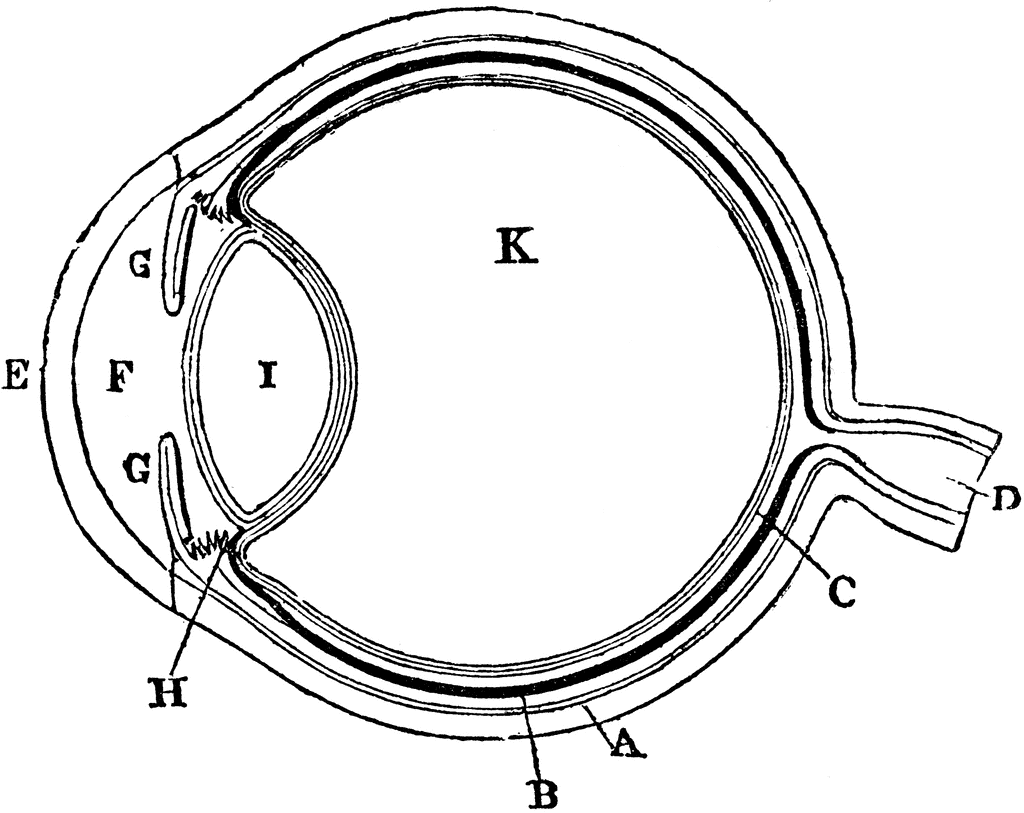

Sheep eye dissection worksheet LAS Assignment 7 - Eye Enucleation Įye cow labeled cows enucleation unlabeled coronal section bio201 return preserved Trusted since 2012, Loopy has 45000+ five star reviews, risk-free returns, and a lifetime warranty Tired of dropping your phone Loopy has a comfy finger. Cow's Eye Dissection - Eye Diagram | Cow Eyes exploratorium Location Of Optic Nerve, Optic Desk, Retina, Macula, Lens, Iris, Pupil eye anatomy diagram eyes label function human vision parts lens eyeball pupil iris cornea optic retina system physiology nerve sclera PPT - COW EYE DISSECTION PowerPoint Presentation - ID:3482425 cow eye dissection optic disc pupil cornea sclera nerve iris retina tapetum powerpoint edu ppt presentation Sheep Eye Dissection Worksheet - Bluegreenish Planarian system digestive platyhelminthes hydra structure internal phylum cavity gastrovascular simple freshwater felina mouth cnidaria Cow Eye Dissection Diagram Labeled | Featuresofthe Cat Eye, Internal dissection labeled dissecting labo Cow Eye Labeled Diagram - ClipArt Best diagram eye labeled cow human eyes system Moderate Energy Dry Cow Diets May Improve Postpartum Health en.ĭry cow dairy cows energy diets requirements moderate cattle far postpartum improve health exceed Required For Module 1. Sheep Brain External View Labeled | Brain Anatomy, Occipital Lobe sheep brain labeled anatomy external dissection lobe physiology frontal nervous system occipital savalli Sheep Brain Dissection Bi - BIOLOGY JUNCTION sheep brain dissection lab ventral inferior anatomy label human bi diagrams companion locate following biologyjunction Freshwater Planarian (Polycelis Felina) - Digestive System 11 Images about Sheep Brain external view labeled | Brain anatomy, Occipital lobe : Cow Eye Labeled Diagram - ClipArt Best, Cow Eye Dissection Diagram Labeled | Featuresofthe Cat Eye, internal and also Freshwater Planarian (Polycelis felina) - Digestive system. Name the three layers you sliced through when you cut across the top of the eye: 4.
COW EYE DIAGRAMS DOWNLOAD
There is a printable worksheet available for download here so you can take the quiz with pen and paper. List four differences between the anatomy of the cow eye and a human eye. This is an online quiz called Cows Eye Diagram.Use the prod to poke a hole in the eye halfway between the cornea and the optic nerve.It may be beneficial to remove some of the fat and/or muscle, as it may be helpful for observation of the Sclera. The fat is a white-yellow tissue and the muscle is the brownish tissue. Notice that it is covered in a layer of fat and muscle. Be mindful and respectful of other students and the task at hand.ĬAUTION: If you get grossed out easily do NOT do this.Be careful of the sharp objects the can cut you or one of the other students.Ensure you discard of all the cow eye parts as instructed by your teacher.Keep your hands away from your mouth and eyes when handling the cows eye.Make sure you wash your hands and all utensils after completing the dissection to ensure that all bacteria has been washed off. Examining the cow eye can help students understand how their own eyes work. What do the human eye and cow eye have in common? There are many similarities and a few differences. This wiki will be created by students from Magee Secondary school with the help of their science teacher, Mr.


In grade 8 science the students dissect an eye. Anterior chamber - the front section of the eyes interior where aqueous humor flows in and out providing nourishment to the eye and surrounding tissues.


 0 kommentar(er)
